Epithelial Morphogenesis
Most experiments have been done around the mouse submandibular gland. The epithelium of the mouse submandibular gland becomes a plastic structure during morphogenesis (Heida, 1996). Cellular plasticity is measured as a function of the structure of the tissue and the amount of adhesion between cells. As the epithelial cells become plastic, movements by the mesenchyme have a large effect on determining epithelial structure, as will be seen later. This plasticity arises at embryonic day 12 period when the epithelium changes from a simple layer into an undifferentiated cell mass by invagination. The mechanics by which the cells become a cell mass are not understood. It seems, though, that epithelial cells have an intermediary state when they are part of this cell mass (Heida, 1996).
The epithelial cells are in a form of cellular limbo as they have characteristics of epithelial cells such as having a surrounding basal lamina and characteristics of mesenchymal cells like a loss of cellular adhesion. This ability to become plastic arises from the loss of certain types of cellular junctions while the epithelial cells are in the cell mass. Cellular junctions like desmoplakins and ZO-1, a tight junction associated protein, disappear in the epithelial cell mass (Heida, et. al, 1996). So in essence the epithelium becomes "de-epithelialized" and is susceptible to the flowing movements of the underlying mesenchyme (Heida et. al., 1996).
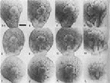
During the next two days the cell mass will differentiate into the branched tree-like form characteristic of a salivary gland. Now, the originally flat epithelium has an apical-basal polarity. There are several methods by which the branching of the salivary epithelium is understood to happen. Strangely enough, one discovery showed that this branching pattern can begin even without the proliferation of the epithelium. Nakanishi (1987) irradiated mouse embryonic submandibular epithelium with X-rays to effectively stop cell division and saw that the initial clefts of the salivary glands still formed (Figure 1). These clefts mark the area where branching will proceed to form the characteristic tree shape of the salivary glands. (Nakanishi, 1987). This proved the assumption that epithelial proliferation initiated branching morphogenesis as well as continued it, incorrect. The many factors that play a part in initiating epithelial cell clefting and branching lay within the basal lamina, like basal lamina glycosaminoglycans and laminin-1 and the mesenchyme.
Figure 1: The effect of X-ray irradiation on the development of the mouse submandibular gland. The top row across represents the control gland at 1 hr., 6, hr., 10 hr., and 27hr. in culture. The bottom two rows demonstrate the effects of radiation levels at 200 rad and 100 rad. There is a significant amount of intial cleft formation in theseglands although cell proliferation has been stopped (Figure from Nakanishi, 1987).
Return to Salivary Gland Contents
Return to Homepage